

Squamous cell carcinoma is the second most common skin cancer, and when it is found early most cases are curable. It usually shows up as a scaly red patch, a firm bump, or a sore that will not heal — often somewhere that has taken years of sun. This post covers what to look for, who is most at risk, and what happens between noticing a spot and finishing treatment.
What It Is
Squamous cell carcinoma starts in the squamous cells that make up the middle and outer layers of the skin. Bakersfield Dermatology's page describes it as usually not life-threatening, but capable of growing large if left untreated and leading to serious complications. Most cases trace back to ultraviolet radiation, from sunlight or from tanning devices.
It is common. The Skin Cancer Foundation estimates 1.8 million cases are diagnosed each year in the United States — about 205 an hour — and reports that incidence has risen by up to 200 percent over the past three decades.
The Warning Signs
Squamous cell carcinoma often appears on sun-exposed skin: the scalp, ears, face and lips. But it can occur anywhere. The practice lists these signs:
- A firm bump or nodule that may be skin-coloured, pink, red, black or brown.
- A flat sore with a scaly crust that does not settle down.
- A new or raised area on an existing scar or sore.
- A rough patch on the lip that may open up.
- A rough or sore area inside the mouth.
- A raised or wart-like sore on or around the anus or genitals.
The Skin Cancer Foundation describes much the same picture — scaly red patches, open sores, rough thickened or wart-like skin, and raised growths with a central depression — and adds that these can crust over, itch or bleed, and tend to persist for weeks.
The single most useful rule: a spot that has not healed after several weeks is worth showing a dermatologist, whatever it looks like. Healing is what ordinary skin does; not healing is the signal.
Who Is Most at Risk
The primary driver is DNA damage from ultraviolet radiation. Risk is higher for people who sunburn easily, who have had a lot of lifetime sun exposure or used tanning beds, who are immunosuppressed, who have had a previous skin cancer, or who have areas of chronic skin inflammation or scarring.
Skin tone changes where to look rather than whether to look. The practice notes that people who sunburn easily tend to find these cancers on sun-exposed areas, while in people with Black and brown skin they may appear in less exposed spots — which is why a proper examination is full-body.
There is also a direct line from precancer to cancer. The Skin Cancer Foundation notes the vast majority of squamous cell carcinomas start off as actinic keratoses, the rough scaly patches that form on chronically sun-damaged skin. Treating those early is the cheapest form of prevention available.
How It Is Diagnosed
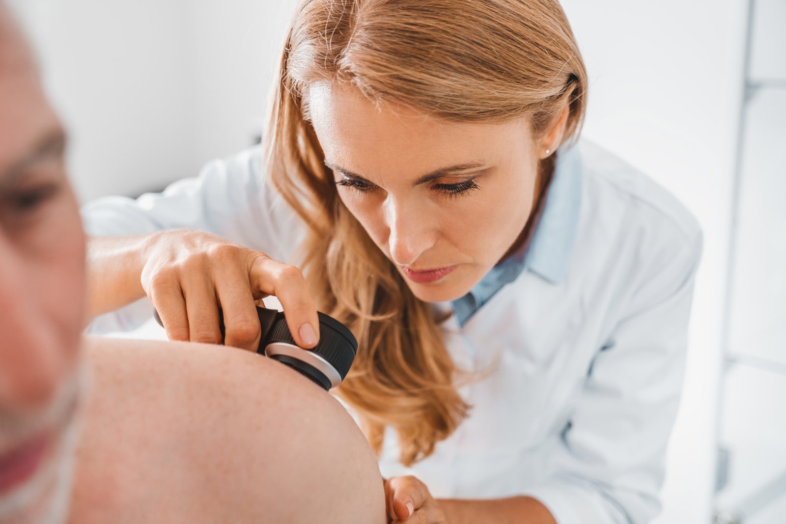
Diagnosis begins with examination, usually with a dermatoscope, and is confirmed with a biopsy — a small sample of the lesion examined under a microscope. Looking alone cannot settle it, because several harmless growths imitate skin cancer and vice versa. A seborrheic keratosis, for instance, is entirely benign but can look alarming.
Treatment Options
What is recommended depends on the size and depth of the tumour, where it sits, and whether it has been treated before. Options offered at the practice include:
- Mohs micrographic surgery: the tumour is removed one thin layer at a time, each layer mapped and examined under a microscope during the visit until no cancer cells remain. It removes the least normal tissue, which matters on the face, ears and other areas where there is little to spare.
- Excisional surgery: the growth is cut out along with a margin of surrounding skin, which is then checked.
- Cryosurgery and electrosurgery: freezing or heat-based destruction, generally for superficial or low-risk lesions.
- Topical therapy: prescription medication applied to the skin, for certain superficial tumours or when surgery is not suitable.
The limitation worth stating: methods that destroy tissue without examining it under a microscope — freezing, for example — cannot confirm how completely the tumour was removed. That is a trade-off your dermatologist will weigh against the size, site and risk of the particular lesion, and the reason follow-up appointments matter after any treatment.
After Treatment
Having one skin cancer raises the chance of finding another, so ongoing skin checks are part of care rather than an optional extra. Between appointments, a monthly skin self-exam is how most new spots get noticed. Population-level statistics like the ones above describe groups, not individuals — your dermatologist will explain what your particular diagnosis means for you.
Get a Spot Examined in Bakersfield
Bakersfield Dermatology & Skin Cancer Medical Group diagnoses and treats skin cancer at 4100 Empire Drive, Suite 120, Bakersfield, CA 93301, with additional offices in Porter Ranch, Valencia and Palmdale. If you have a sore that has not healed or a growth that is changing, call 866.903.3376 or request an appointment for a skin examination.
Frequently Asked Questions
How fast does squamous cell carcinoma grow?
It varies. Some stay small for months, others enlarge over weeks. The Skin Cancer Foundation notes that untreated squamous cell carcinoma can grow into deeper layers of skin and spread, which is why a sore that has not healed after several weeks should be examined rather than watched.
Does a squamous cell carcinoma always start as an actinic keratosis?
Not always, but there is a strong link. The Skin Cancer Foundation notes that the vast majority of squamous cell carcinomas begin as actinic keratoses, which is the reason those rough precancerous patches are treated early.
Can I get it on skin the sun does not reach?
Yes. Most cases come from UV exposure, but the practice's page notes it can occur anywhere, including inside the mouth and on the genitals, and that in people with Black and brown skin it may appear in less sun-exposed areas. That is one reason a full-body exam covers more than the obvious places.
Will treatment leave a scar?
Any procedure that removes tissue leaves a mark. Mohs micrographic surgery is used partly because it removes the least normal tissue around the tumour, but the size and location of the growth still shape the result. Your dermatologist can tell you what to expect for your particular spot.
Sources: The Skin Cancer Foundation, Squamous Cell Carcinoma (updated September 2026) — 1.8 million annual cases, the rise in incidence, warning signs, risk factors, the link to actinic keratosis and early-treatment outlook; and Bakersfield Dermatology's own Squamous Cell Carcinoma page — description, symptom list and the treatments offered here.